
Heel hook injuries in rock climbing occur due to excessive force around the knee joint. Since a heel hook requires a bend, an externally rotated knee, and a high load, it makes structures like the lateral meniscus, biceps femoris muscle, and medial collateral ligament prone to injury.
In this blog, I’ll discuss how a heel hook injury occurs, which structures may get injured and how, and what you can do to prevent heel hook injuries when rock climbing or bouldering.
Are you ready?
Let’s dive in!
1. How Do You Get an Injury from a Heel Hook?
The heel hook is a climbing-specific movement in which you use your heel to stabilize yourself on rock or on the climbing wall. For optimal control, you bend your knee and rotate the lower leg out so you can press your heel better into the wall.
If this specific heel hook movement is done in combination with one or more of the following:
- maximal force output
- sudden movement changes like slipping off a hold
- lack of solid warming-up
- repetitive heel hooking without the appropriate training
you will be more likely to get injured. The high amount of force emanating from the knee is bent and rotated outwards generating large stresses on the medial collateral ligament (MCL), the lateral collateral ligament (LCL), the lateral meniscus, the iliotibial band (IT-Band), and the biceps femoris and popliteus muscles.
2. Which Structure Can Get Injured from a Heel Hook?
The LCL, MCL, lateral meniscus, IT-Band, and the biceps femoris and popliteus muscles are the structures that can be injured as a result of a heel hook. It depends on your physical condition and the type of heel hook which structured might get injured first.
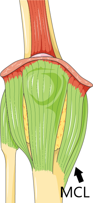
2.1 Medial Collateral Ligament (MCL)
The MCL is on the inside of the knee, originates on the femur, and connects to the tibia. It prevents excessive valgus. This is a sideways movement, where the lower leg moves laterally as opposed to the upper legs. To give you an idea of what that looks like, “X-legs” or knocking knees come with excessive valgus in de knee joint.
Thus, if your heel hook generates too much valgus force, which is more likely with the flexion and external rotation of the knee, your MCL might tear.
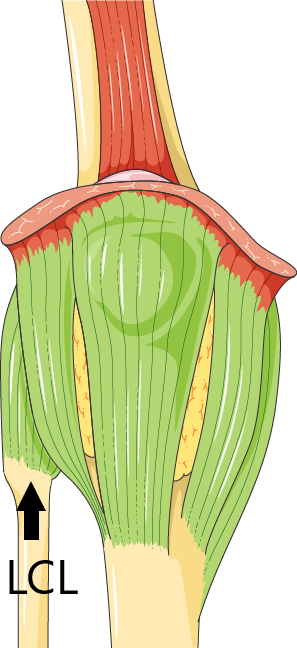
2.2 Lateral Collateral Ligament (LCL)
The LCL is on the outside of the knee, originates on the femur, and connects to the fibula. It has the exact opposite function of the MCL and thus prevents excessive varus or “O-legs”.
If you place a heel hook that provokes too much varus your LCL might tear.
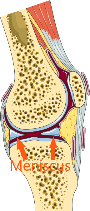
2.3 Lateral Meniscus
The two half-moon-shaped menisci in your knee help absorb impact forces and enhance rotation. When you internally rotate your knee there’s more pressure on your medial meniscus and when you externally rotate there’ll be more pressure on the external meniscus.
Consequently, every heel hook puts high pressure on the lateral meniscus and when this pressure becomes too high your meniscus might tear.
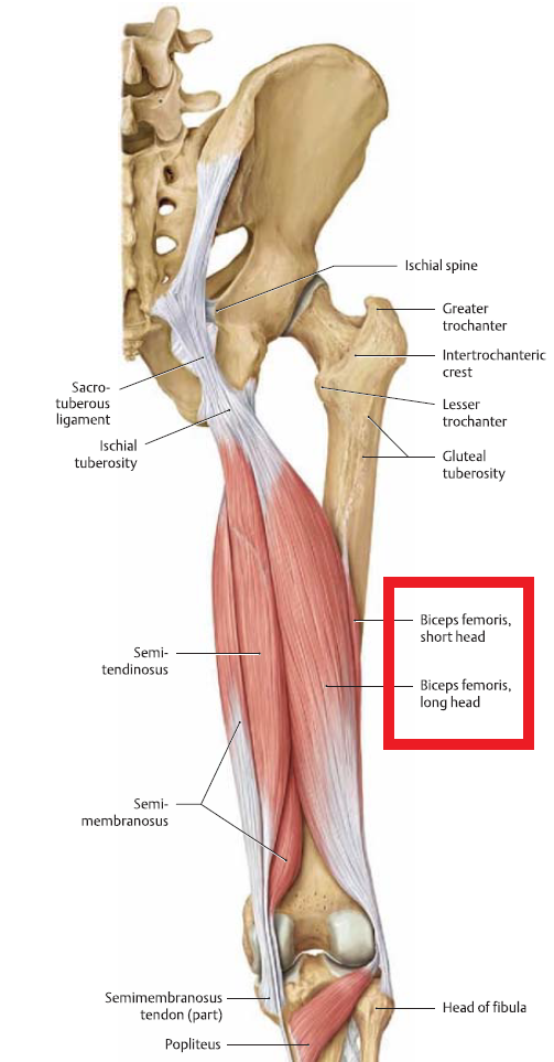
2.4 Biceps Femoris Muscle
The biceps femoris muscle consists of 2 parts, the biceps femoris caput longum (long head) and caput Brevis (short head). Together with the semitendinosus and semimembranosus muscle, it makes up the hamstring muscle group. The biceps femoris is on the lateral side of the leg and thus has to work hardest when performing heel hooks.
Repetitive microtrauma, repetitive stresses, and sudden slips of the heel could lead to strains or tears of the biceps femoris muscle.
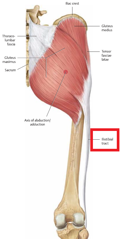
2.5 Iliotibial Band (IT Band)
The IT Band is a tendonous plate that originates at the hips through the gluteus maximus and tensor fascia lata muscles and connects to the fibula with LCL. Since the hip, just like the knee, needs to externally rotate as well while doing heel hooks the IT Band has to deal with a lot of tension too.
You might suffer acute injuries of the IT Band, but you could also experience pain along with your glutes, and/or at the lateral side of your upper leg and knee due to hypertension of the IT Band.
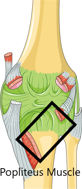
2.6 Popliteus Muscle
The popliteus is a small muscle in the back of the knee that serves as an internal rotator. When performing a heel hook you lengthen the popliteus to get into external rotation. This means the popliteus has to work in its weakest position since the closer a muscle is to its full length the harder it is to generate force. And the harder it is to generate force, the more likely the muscle is to be overloaded. This could either lead to hypertension and pain or to an acute injury involving a strain or tear of the muscle fibers.
3. Heel Hook Injury Treatment
The treatment of heel hook injuries depends entirely on the injured structure and the severity of the injury. When a ligament, muscle, or tendon is completely torn, surgery is an option. A meniscus tear might or might not heal by itself and surgery, in this case, might be an option too. In all other situations, you can heal your injury conservatively, preferably with the help of a rehab professional.
The best way to deal with heel hook injuries though is to never get one in the first place.
4. How to Prevent a Heel Hook Injury
The best ways to prevent heel hook injuries are:
- Building your exposure to heel hooks slowly: start with heel hooks on big holds progressing to small holds. And progress from vertical to overhanging terrain.
- Use the minimally necessary amount of tension to hold the heel hook
- Practice heel hooks often without the stress of trying to send a project. Work on your technique and develop a feeling for what a solid heel hook feels like. This way you’ll notice when you are not hooking right and are exposed to a higher risk of injury.
- Stretching increases your muscles and connective tissues’ resistance to stretch and might prevent them from tearing in gnarly positions.
- Train your legs(!). With stronger leg muscles you can handle more force during heel hooks. Think of exercises like the glute-ham raise, deadlifts, and ATG Split Squats.